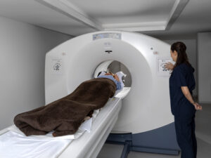
 In June 2024, we provided an overview of the different types of imaging scans you may receive if you have MBC. Then we began a deeper dive into each scan, starting with MRI scans in July 2024. We now continue our series by highlighting positron emission tomography (PET)/computed tomography (CT) scans.
A PET scan is an imaging test that uses a radioactive substance called a tracer that can look for and attach to cancer cells. Two commonly used tracers are fluorodeoxyglucose (FDG) and fluoroestradiol (FES). A CT scan produces body pictures created by X-ray energy. These two scans are often combined. A PET/CT scan can see sites of metastasis throughout the body, although an FES PET/CT scan cannot be used to look for cancer in the liver.
Participating in a clinical trial that is studying imaging scans may be an opportunity to get a scan you might otherwise not get because of considerations such as insurance or a non-standard use for the scan.
Read below to learn more about what CT and PET scans are, how PET/CT scans are used in MBC, and for clinical trials studying these scans in people with MBC.
Introduction to CT Scans and PET Scans
- American Cancer Society: A CT scan, also called a CAT scan, uses X-rays to create a detailed picture of the inside of the body
- Breastcancer.org: PET scans use a radioactive tracer to create images of places in the body where cancer may be
PET/CT Scans for People with MBC
MBC Clinical Trials
- Metastatic Trial Search: Trials for Imaging
Last Modified on September 3, 2024
